Lengthy wait times for medical diagnostic imaging not only delay critical procedures, they add additional strain to patients and families who are left waiting in uncertainty.
On average, Ontarians wait 107 days while Windsorites wait 102 days for a Magnetic Resonance Imaging (MRI) scan, according to the Ontario Ministry of Health and Long-term Care.
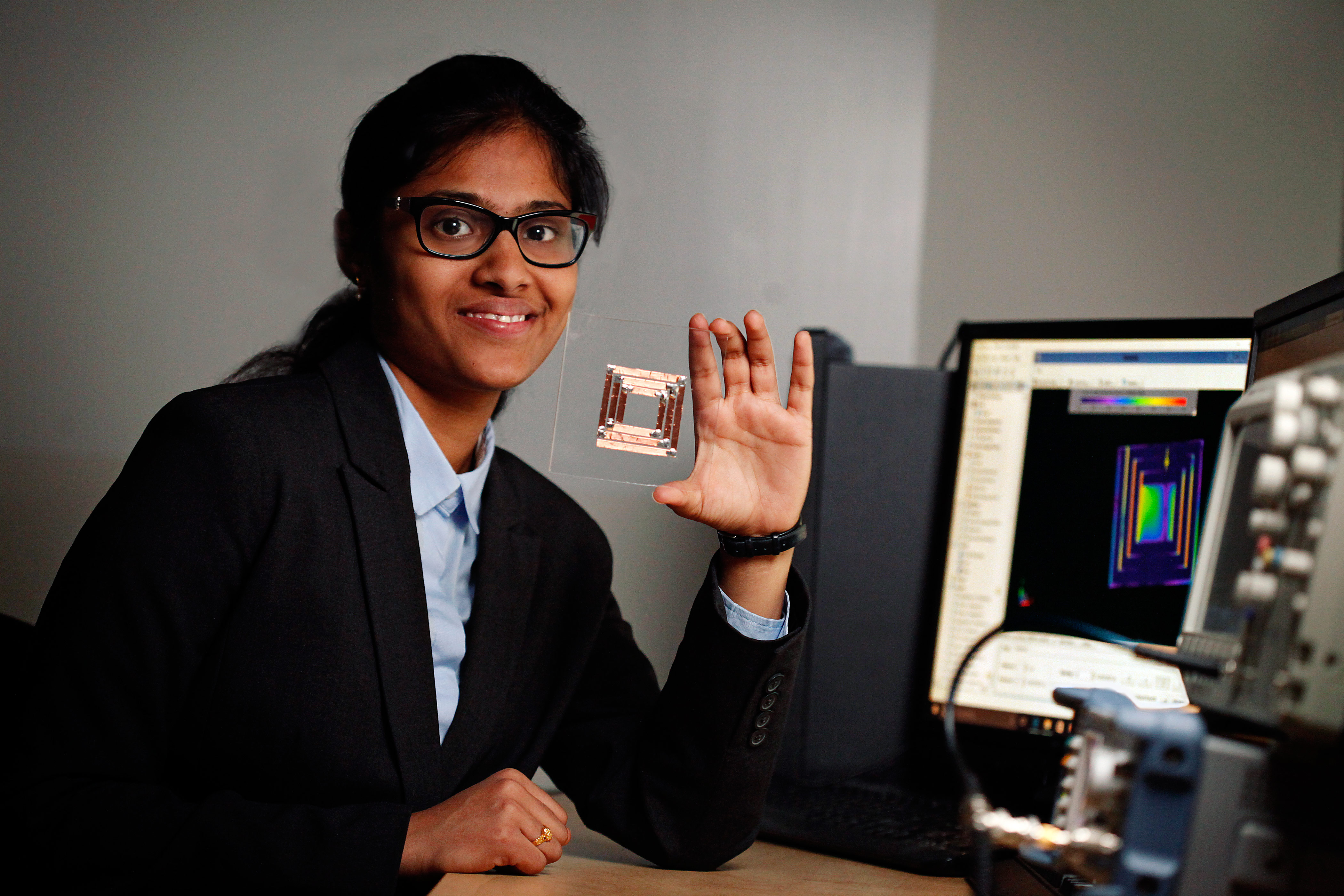
A University of Windsor graduate student is hoping her promising new research can prove to be a game changer for the medical field and the thousands of people waiting in limbo for critical information.
Sujitha Vejella, an electrical and computer engineering master's candidate, has designed a microelectromechanical system (MEMS) based radar for medical diagnostic imaging under the supervision of Dr. Sazzadur Chowdhury, an associate professor of electrical engineering. Using MEMS, a technology that enables the creation of 3D microscopic devices with moving parts, Vejella has fashioned a portable and low-cost imaging system roughly the size of a Rubik’s Cube, offering an alternative to MRI machines, which are expensive and not easily accessible.
“It’s safer than CT and X-Ray scans and there are no side effects,” said Vejella, who is working on completing a 16 by 16 pixel prototype. “We can use this technology to detect cancer at an early stage and determine the tumor size and location.”
Vejella is among nine finalists who will compete in the Microsystems Design Award category at the TEXPO Competition held by CMC Microsystems — a network that supports researchers and industry across Canada’s National Design Network. The graduate student competition will be held on Oct. 17 in Montreal at Innovation 360, the CMC Microsystems annual conference that brings together Canadian micro-nano innovators from academia, industry and government.
Vejella’s design uses an ultra-wideband radar to generate a pulse which is reflected back in layers, generating a 3D image of the targeted organ using arrays of small microwave pixels without ever coming into contact with the patient.
“This produces a high resolution image compared to conventional imaging systems at a much lower cost,” said Dr. Chowdhury.
The technology can also monitor a patient’s vital and cardiac signs and alert healthcare professionals of critical changes. Although she is in the early stage of her research, Vejella will have to demonstrate industrially relevant research results and will be evaluated on presentation and technical excellence, application to industry and visual effectiveness. In addition to receiving complimentary travel and accommodation, she is competing for a $3,000 award that can be used to support education, training, conference participation or lab visits related to microsystems.
Last year, Rayyan Manwar, a UWindsor, electrical and computer engineering, PhD student, finished in second in the TEXPO Huawei Microsystems Design Award category for his research in MEMS.