Functional Near Infrared Spectroscopy (fNIRS) refers to the use of near-infrared spectroscopy (NIRS) for functional imaging. The SRCC, in collaboration with researchers from Dr. Abeare's lab, has begun using fNIRS to image brain activity after concussion.
Why fNIRS?
In typical functional neuroimaging (e.g., fMRI), there is a tradeoff between spatial resolution (being able to tell where a neural signal is coming from) and temporal resolution (being able to tell when a neural signal is occurring). That is to say, if you have a good idea of where a neural signal is coming from, you're often measuring too slowly. The technology behind fNIRS allows it to have high spatial and temporal resolution. Additionally, fNIRS measurements can be taken while moving, unlike fMRI, where the patient has to be completely still during measurement.
How does fNIRS work?

When neurons fire during brain activity, this causes responsive vasodilation - blood vessels nearby dilate. This increased bloodflow at the site of the active brain area can be measured as a proxy for neural activity (such as in fMRI). Incoming blood is oxygenated, and outgoing blood is deoxygenated. fNIRS uses near-infrared lights to detect changes in oxy- and deoxyhemoglobin in the blood, because oxygenated and deoxygenated blood have different levels of light absorption.
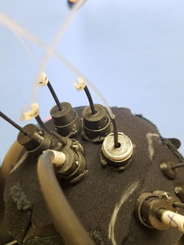
fNIRS uses a set of near-infrared lights (sources) and sensors (detectors) attached to the head designed in an organized array called a montage. Sources and detectors are arranged in such a way that traingulation can be used to determine the location and depth of a given neural signal (spatial resolution < 1cm). Analysis of the data and 3D reconstruction results in time-sensitive heatmaps of activity.
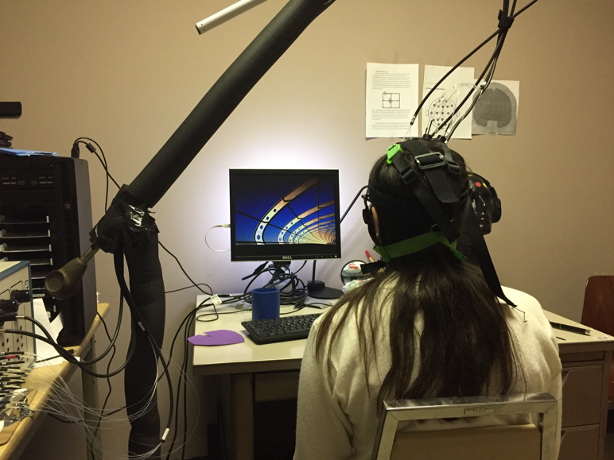
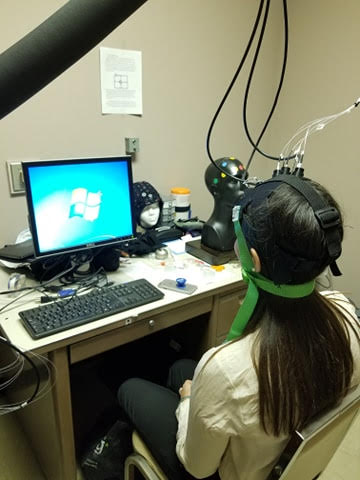
Example of an fNIRS set up:
Example of a montage and the corresponding locations on the brain of sources (blue) and detectors (red):