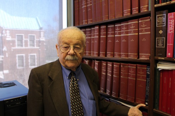
 Mordechay Schlesinger, physics professor and co-inventor of the non-invasive diagnosis technique that monitors tumour activity to confirm if chemotherapy or radiation therapy is working.
UWindsor researchers have patented a non-invasive diagnosis technique that monitors tumour activity to confirm if chemotherapy or radiation therapy is working.
“In the case of chemotherapy, you are poisoning the person and the tumour,” says Mordechay Schlesinger, physics professor and co-inventor of the technique.
“You are poisoning the healthy cells along with the cancerous ones, all in hopes that the tumour dies before the person. Therefore it is crucial to check that the treatment is working, because unfortunately, it is too often that the chemo kills the person before the cancer does.”
The method was invented by Dr. Schlesinger and his post-doctoral student Long Jian Liu (MSC 2009, PhD 2013), along with John R. Ewing and Steven L. Brown, two medical physicists from Henry Ford Hospital in Detroit. The patent is titled, Method and Apparatus for Diagnosis of Tumor Activity Using Tumor Interstitial Fluid Pressure.
“The exciting part is that this means the U.S. patent office has officially recognized this method as a unique, practical and worthwhile practice,” Schlesinger says.
A group of scientists from Oslo University Hospital’s Institute for Cancer Research, who are using the technique, published a paper on the method in the academic journal, Cancer Research.
The Oslo University Hospital paper also cited Schlesinger’s team’s paper, Phenomenological model of interstitial fluid pressure in a solid tumor, from the journal, Physical Review E.
The team’s imaging process documents the interstitial fluid pressure (TIFP) of the cells in the tumour. Schlesinger says pressure differentiates between cancer cells and healthy cells, so documenting pressure change shows if chemotherapy or radiation therapy is killing the tumour’s cells.
Schlesinger says his team’s method uses a scanning apparatus to make multiple images of a tumour, which is used in conjunction with a dye that acts as a visual marker to show a rate of change resulting from chemotherapy.
Until now, using a needle to monitor pressure was the only way to determine if chemotherapy or radiation therapy was giving the desired results.
“Depending on the type of cancer, this test couldn’t even be done,” Schlesinger says.
“When it comes to something like a tumour in your brain, using a needle is out of the question.”
He says this new technology is a game changer in cancer therapy and promises that work will continue to refine the technique. As part of the patent, the research team will work to modify Magnetic Resonance Imaging (MRI) machines to allow the operator to determine tumour pressure without much difficulty.
“It is great news that Oslo is already using the method and they’ve learned it is most useful for women with ovarian cancer.”
Schlesinger says any royalties received from this patented process will be returned to the University of Windsor and Henry Ford Hospital.
“I’m not as young as I used to be. This thing is for each and every one of us because we are all candidates for cancer. Nobody can say I will never have cancer, so ultimately I’m helping myself.”